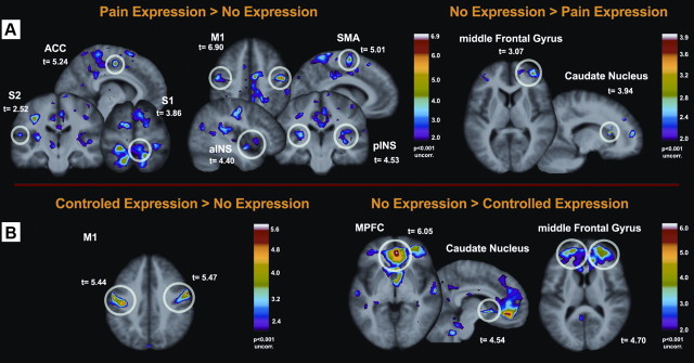
Figure 3.
Within-subject analyses—differences between painful events with and without facial expressions of pain. A, Stronger brain activation was found in M1 (face area) and in several pain-related areas in trials where individuals displayed spontaneous pain expression (left). In contrast, stronger activation was found in prefrontal areas and caudate nucleus when no expression was displayed (right). B, In facially non/low-expressive individuals, the display of intentionally communicated facial expressions of pain was associated with increased activity in M1 (left) and reduced activity in prefrontal cortices and in the caudate nucleus (right). *Directed search threshold at T = 2.84 (p = 0.05). Bonferroni-corrected for 12 pain-related regions.

