
Figure 1.
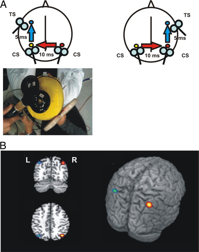
A, Schematic representation of the experimental procedure. TMS of the primary motor cortex (M1) evokes a small twitch in contralateral hand muscles that can be measured with surface electromyography. At rest, this response is enhanced following ipsilateral PPC stimulation, implying the activation of a corticocortical pathway that originates in PPC (Koch et al., 2007) and thus indicating the activation state of PPC (blue arrows). To investigate the interhemispheric interactions between the right and left PPC (red arrows), we developed a trifocal stimulation method. TMS of right PPC preceded TMS of left PPC by 10 ms. A third stimulus was applied 5 ms later on the ipsilateral left M1. The same experimental scheme was reversed to test the effects of left PPC over the right PPC (bottom). B, Anatomical location of right (R, red/yellow) and left (L, green/blue) PPC TMS sites in MNI coordinates. The colored spots represent the mean group coordinates (brighter color) ± SD (darker outline).