
Figure 8.
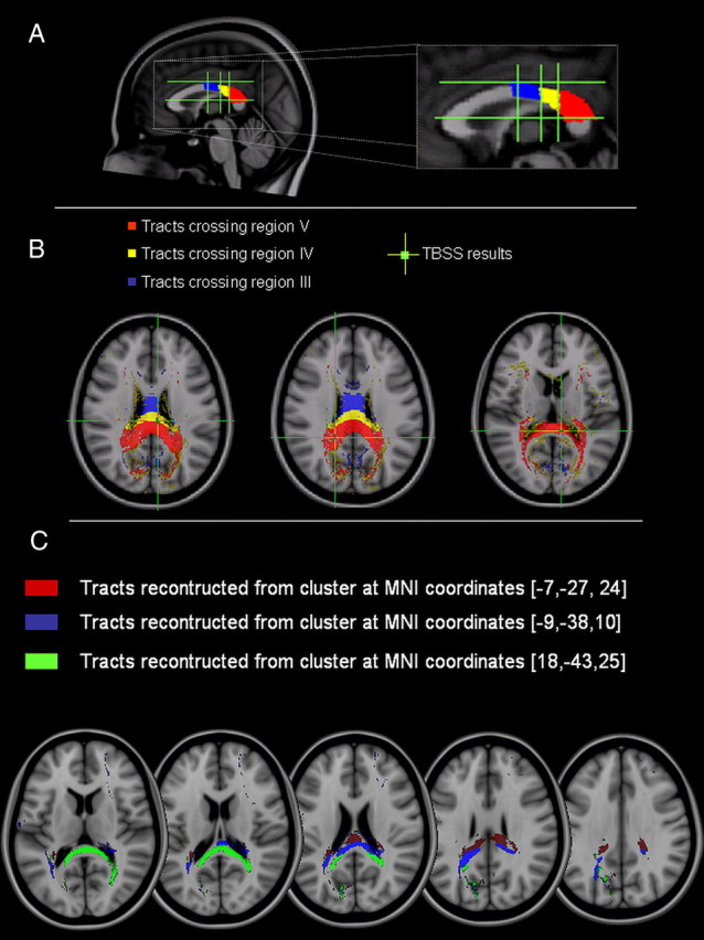
Tractography of the posterior portion of the CC. A, Segmentation of the midsagittal slice according to the topography of the CC proposed by Hofer and Frahm (2006). B, Reconstruction of CC fibers crossing regions III (blue), IV (yellow), and V (red). The green crosses localize the areas of significant correlation obtained with the TBSS analysis between the individual changes of the left PPC–M1 connection following TMS of the right PPC at 70% RMT and FA. These areas are localized in either region IV or V. C, Pathways reconstructed using the three CC clusters obtained with the TBSS analysis.