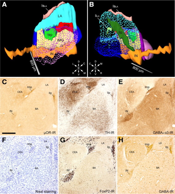
Figure 1.

Full 3D reconstruction and immunohistochemical profile of the ITC clusters of the mouse amygdala. A, B, Medial (A) and lateral (B) views of ITC clusters, CEA (CEc/l in purple; CEm in light purple), and BLA (LA in light blue and BA in blue) of the mouse amygdala. The IN (orange), located ventromedially to the BA, extended rostrally beneath the anterior commissure for considerable length. Rostrocaudally along the intermediate capsule, we observed two discontinuous clusters: the Iap (green) and Imp (red). The intermediate capsule interposed between the Iap/Imp and the IN was identified as IMG (yellow) (de Olmos, 1990). The clusters observed laterally and dorsally to the BLA, previously known as paralaminar nucleus (Amaral and Price, 1984), showed no apparent cellular continuity in mouse and were thus named Ilp (dark green) and supralateral cluster (IsLA, pink), respectively. A small cluster inside the LA was also observed and termed intralateral cluster (IiLA, light green). C–H, Representative micrographs of consecutive sections (bregma levels, −1.46 to −1.70) of the IN, Imp, and Ilp visualized through different immunohistochemical stains: μOR-IR (C), TH-IR (D), GABAA α3-IR (E), cresyl violet (F), FoxP2-IR (G), and GABA-IR (H). Despite each ITC cluster receiving a relatively strong catecholaminergic innervation (TH-IR), as reported previously in rat (Fuxe et al., 2003; Muller et al., 2009), we observed that the caudal half of the IN (D) and the most caudal portion of Imp and Ilp were almost devoid of TH-IR. BA, Basal nucleus of amygdala; CEA, central nucleus of amygdala; Iap, anterior paracapsular ITC cluster; IiLA, intralateral paracapsular ITC cluster; Ilp, lateral paracapsular ITC cluster; IsLA, supralateral ITC cluster; LA, lateral nucleus of amygdala; L, lateral; D, dorsal; C, caudal; R, rostral; V, ventral; M, medial. Scale bars: A, B, 800 μm; C–H, 250 μm.
