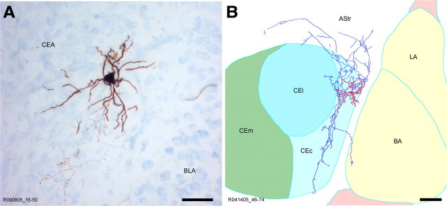
Figure 3.
Morphological features and axonal pattern of a representative CEc neuron. A, B, Light microscopy micrograph (A) and reconstruction by camera lucida (B) of a representative in vitro recorded and biocytin-filled CEc neuron. The soma and dendrites are labeled in red, whereas the axon is marked in blue. The boundaries of the amygdaloid subregions have been drawn referring to the section containing the soma. AStr, Amygdalostriatal transition area; LA, lateral nucleus of amygdala. Scale bars: A, 50 μm; B, 100 μm.

