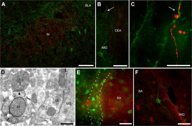
Figure 7.
mGlu1α receptor-immunopositive cells encircle ITC clusters and are targeted by MCp–Imp neurons. A, Double-immunofluorescence micrograph depicting IN clustered neurons immunolabeled for FoxP2 (in red) encircled by the soma and dendrites of large neurons immunolabeled for mGlu1α receptor (in green). B, C, Low-power (B) and high-power (C) micrograph of the filled axon (in red) of a MCp–Imp recorded neuron potentially forming a synaptic contact (arrow) onto a mGlu1α receptor-immunopositive dendrite (in green) within the intermediate capsule. D, Pre-embedding immunoelectron microscopy confirmed that the observed contact is indeed a symmetric synapse (arrowhead). In the ITCs, the soma of neurons containing mGlu1α receptors do not express GABA as revealed by the lack of colabeling between mGlu1α receptor (in red) and GFP (in green) in GAD65-GFP transgenic mice (E) or between mGlu1α receptor (in green) and GABA (in red) (F). ld, Labeled dendrite; lt, labeled terminal. Scale bars: A, 100 μm; B, 50 μm; C, 15 μm; D, 500 nm; E, F, 50 μm.

