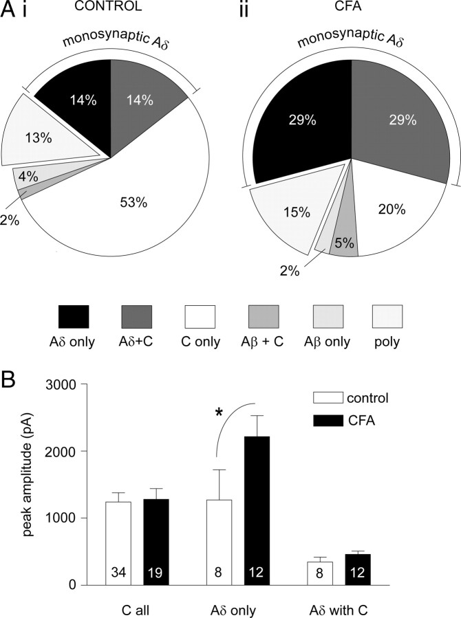
Figure 5.
CFA inflammation increases the incidence and magnitude of monosynaptic Aδ-fiber but not monosynaptic C-fiber input to lamina I NKIR+ neurons. A, The proportion of lamina I NK1R+ neurons with input from different afferent fiber classes in both control (Ai) and CFA inflammation (Aii) groups is summarized (control neurons, n = 56; CFA neurons, n = 41). The wedges in the pie chart refer to the primary afferent monosynaptic input(s) received apart from the “poly” grouping, which reflects those neurons receiving purely polysynaptic inputs. For example “Aδ only” refers to those neurons receiving only monosynaptic Aδ-fiber input, and “Aδ with C” refers to those neurons receiving both monosynaptic Aδ-fiber and monosynaptic C-fiber input. The overall pattern of afferent input is significantly altered by CFA inflammation (χ2 analysis, p < 0.05). The incidence of monosynaptic Aδ-fiber input is significantly increased by twofold (Fisher's exact test, p < 0.01), whereas the incidence of C-fiber monosynaptic input is not significantly affected (Fisher's exact test). B, Effect of CFA inflammation on the peak amplitude of monosynaptic Aδ-fiber-evoked and monosynaptic C-fiber-evoked EPSCs in lamina I NK1R+ neurons. “C all” refers to the C-fiber-evoked peak amplitude in all neurons receiving monosynaptic C-fiber input, “Aδ only” refers to the Aδ-fiber-evoked peak amplitude of those neurons receiving only monosynaptic Aδ-fiber input, and “Aδ with C” refers to the Aδ-fiber-evoked peak amplitude of those neurons receiving both monosynaptic Aδ-fiber and monosynaptic C-fiber input. Averaged data are represented as mean ± SE, and the numbers of neurons analyzed within each class were shown on each bar. Two-way ANOVA followed by Bonferroni's post hoc tests (*p < 0.05) reveals that CFA inflammation significantly increases the peak amplitude in the “Aδ only” grouping.

