Figure 6.

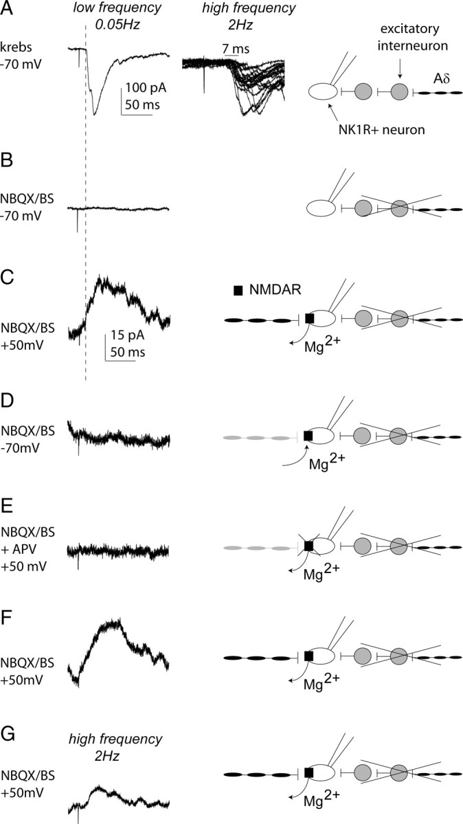
Silent monosynaptic Aδ-fiber input to a control lamina I NK1R+ neuron. A, Characterization of primary afferent synaptic input to a control lamina I NK1R+ neuron receiving polysynaptic Aδ-fiber input. In A–F, the left trace shows examples of EPSCs evoked by stimulation (0.1 ms) using Aδ (100 μA)-fiber stimulation intensities at low frequency. Each trace comprises three averaged traces evoked at 0.05 Hz. The second trace in A shows examples of EPSCs evoked by higher-frequency stimulation (100 μA/2 Hz) and comprises 20 superimposed traces exhibiting a latency variability of 7 ms and is shown at a faster timescale than the left trace. In A–G, the schematic on the right illustrates the input recorded under the different experimental conditions. Scale bars for the left trace in A also apply to B. In B–G, all recordings are performed in the presence of NBQX (10 μm) to block polysynaptic activity and bicuculline (B; 10 μm) and strychnine (S; 1 μm) to block inhibition. B, Stimulation of the neuron, held at −70 mV under these conditions, produced no detectable EPSC, and the block of polysynaptic input is shown schematically. C, When the neuron is held at +50 mV, a slow outward current was observed because of the removal of the voltage-dependent Mg2+ block of the NMDA receptor and unmasking of a silent monosynaptic Aδ-fiber input, as illustrated. Note that the slow outward current has a shorter latency than the polysynaptic input in A (see dotted line). D, The slow outward current was no longer evident on return to −70 mV because of the voltage-dependent Mg2+ block of the NMDA receptor and remasking of the silent monosynaptic Aδ-fiber input, as depicted. E, The outward current was completely blocked by APV (30 μm) as expected for a pure NMDA receptor synapse; this block was reversible (F), and these are shown schematically. In G, the trace comprises 20 averaged traces evoked by 2 Hz Aδ (100 μA)-fiber stimulation intensity (0.1 ms), and the silent monosynaptic Aδ-fiber input is again illustrated. Scale bars in C apply to C–G.
