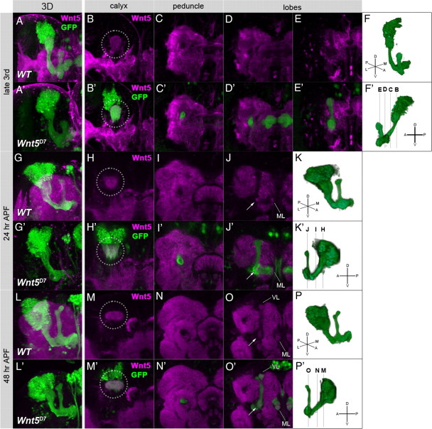
Figure 5.
The expression pattern of Wnt5 in the brain. The Wnt5 protein (magenta) was broadly expressed in the brain at the late third larval instar (A, B–E′), 24 h APF (G, H–J′), and 48 h APF (L, M–O′). The signal of Wnt5 was abolished in the brains of Wnt5 mutants at the late third larval instar (A′), 24 h APF (G′), and 48 h APF (L′). Three-dimensional reconstructed images (A, A′, G, G′, L, L′) and sections at the level of the calyx (B, B′, H, H′, M, M′), the peduncle (C, C′, I, I′, N, N′), and the lobes (D–E′, J, J′, O, O′) are indicated. The structure of the MB was visualized by the expression of GFP (green) driven by OK107-Gal4. The level of each section is schematically illustrated in lateral views of the MB at each stage (F′, K′, P′). Wnt5 was localized in the calyx (B, B′, H, H′, M, M′, dotted circles) at all stages. At 24 h APF, Wnt5 was also detected at the branch point of the lobes (J, J′, arrow) and at the tip of the medial lobe (J, J′, ML). At 48 h APF, Wnt5 was accumulated at the branch point of the lobes (O, O′, arrow) and at the tips of both the vertical and the medial lobes (O, O′, VL and ML).

