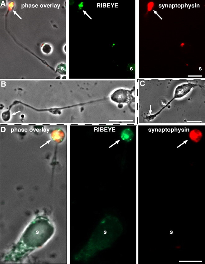
Figure 1.

Dissociated mouse retinal bipolar cells. A–D, Characterization of isolated mouse retinal bipolar cells by immunocytochemistry (A, D) and phase contrast microscopy (B, C). Acutely dissociated (A, B) and 2 d cultured (C, D) mouse retinal bipolar cells were analyzed by immunocytochemistry (A, D) with antibodies against RIBEYE and synaptophysin as indicated. s, Soma of retinal bipolar cells. The arrows denote the presynaptic terminals of retinal bipolar cells that contain RIBEYE-labeled synaptic ribbons and synaptophysin-labeled synaptic vesicles that fill the entire presynaptic terminal. Scale bars: 10 μm.
