Figure 3.
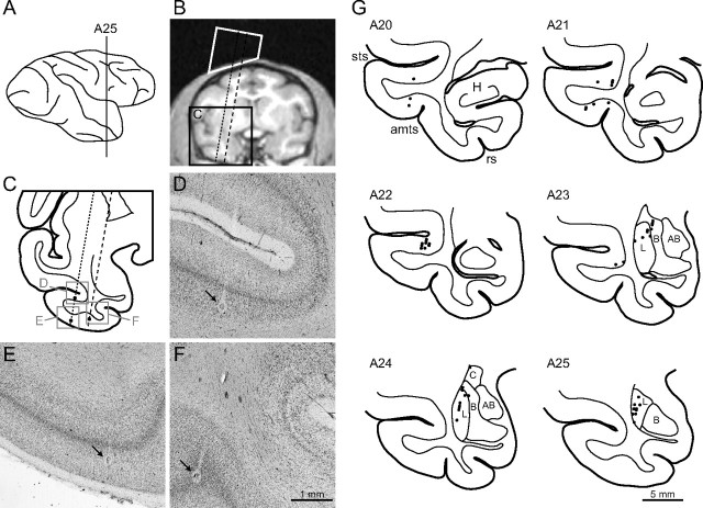
Recording sites. A, Lateral view of the right hemisphere of Monkey S. The vertical line is positioned at 25 mm anterior to the ear canal (A25). B, A coronal slice of magnetic resonance images at A25. The white trapezoid indicates the recording chamber. Dotted and dashed lines indicate electrode penetrations to the temporal cortex and the amygdala, respectively. C, A coronal section corresponding to a region surrounded by the black rectangle in B. D–F, Photographs of regions corresponding to the gray squares in C. Arrows indicate electric lesions made along the penetrations. G, Reconstructed recording sites in the right hemisphere of Monkey S. H, Hippocampus; L, lateral nucleus; B, basal nucleus; AB, accessory basal nucleus; C, central nucleus of amygdala; sts, superior temporal sulcus; amts, anterior middle temporal sulcus; rs, rhinal sulcus. The SI values did not differ in the lateral (n = 21) and basal nuclei (n = 6) of the amygdala in Monkey S (Mann–Whitney test; p = 0.66).

