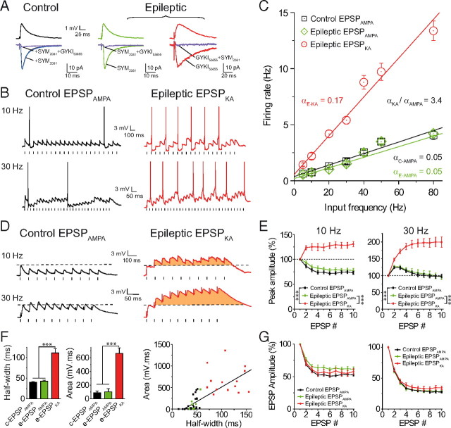
Figure 1.
EPSPKA but not EPSPAMPA generate a sustained discharge in epileptic dentate granule cells. A, Average EPSPs (top) and EPSCs (bottom) recorded in control and epileptic dentate granule cells. Recordings were performed in the presence of gabazine (5 μm), CGP (5 μm), and d-APV (40 μm). Control EPSC before (black trace), after 10 μm SYM 2081 (blue trace), and after 10 μm SYM 2081 + 10 μm GYKI 53655 (purple trace). Epileptic EPSC with 10 μm SYM 2081 (green trace), and with 10 μm SYM 2081 + 10 μm GYKI 53655 (purple trace); epileptic EPSC with 10 μm GYKI 53655 (red trace), and with 10 μm GYKI 53655 + 10 μm SYM 2081 (purple trace). B, Spike discharge for control EPSPAMPA and epileptic EPSPKA at 10 or 30 Hz in DGCs. C, Input/output relationship (α = slope) for EPSPAMPA and EPSPKA (correlation coefficient r = 0.99) showing a multiplicative gain change of input–output operation for EPSPKA. D, Summation of control EPSPAMPA and EPSPKA evoked at 10 and 30 Hz. Note the EPSPKA-mediated depolarizing envelope in orange. E, Summation profile of EPSPAMPA and EPSPKA in control and epileptic conditions, evoked at 10 or 30 Hz. F, Half-width of EPSP and charge transfer of depolarizing envelope for control (c-EPSPAMPA), and epileptic EPSPs (e-EPSPAMPA and e-EPSPKA), and their correlation (r = 0.74). G, Short-term depression of EPSPs at 10 Hz (left) or 30 Hz (right). In this and following figures, spikes are truncated and electrical stimulations are represented by small dashes below the traces; *p < 0.05, **p < 0.01, ***p < 0.001.

