
Figure 1.
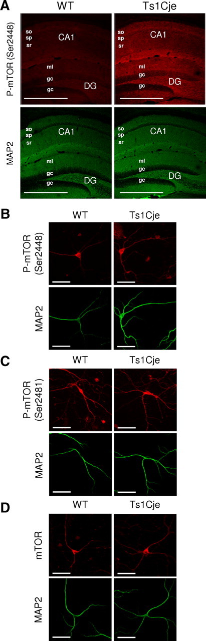
mTOR is hyperphosphorylated in Ts1Cje hippocampus. A, Immunohistochemical detection of phospho-mTOR (Ser2448) protein showing an increased signal in the dendritic layers (identified as MAP2 positive) of the Ts1Cje when compared with the WT adult hippocampus. The maximal projection of a stacking of confocal images is shown in each case. Scale bars, 600 μm. so, Stratum oriens; sp, stratum pyramidale; sr, stratum radiatum; DG, dentate gyrus; ml, molecular layer; gc, granule cell layer. B–D, Representative immunocytochemistry images of phospho-mTOR (Ser2448), phospho-mTOR (Ser2481), and mTOR (total protein), respectively, in WT and Ts1Cje hippocampal neurons at DIV12. MAP2 was used as a dendritic marker. Scale bars, 60 μm.