
Figure 5.
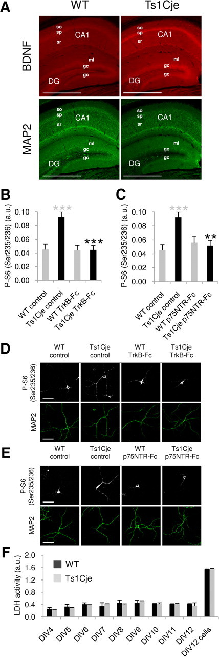
Basal BDNF levels are increased in the Ts1Cje hippocampus. A, Immunohistochemical detection of BDNF proteins (mature and pro-BDNF) showing an increase in the dendritic layers (identified as MAP2 positive) of Ts1Cje versus WT adult hippocampus. Stronger BDNF expression in somatic layers (stratum pyramidale and granule cells) is also appreciable. The maximal projection of a stacking of confocal images is shown in each case. Scale bars, 600 μm. so, Stratum oriens; sp, stratum pyramidale; sr, stratum radiatum; DG, dentate gyrus; ml, molecular layer; gc, granule cell layer. B, Quantification of phospho-S6 (Ser235/236) fluorescence intensity in dendrites of WT and Ts1Cje hippocampal neurons at DIV12, in the presence or absence of the BDNF scavenger TrkB-Fc (1 μg/ml for 24 h), as indicated. A significant decrease was observed in TrkB-Fc-treated versus untreated Ts1Cje cells (n = 9; ***p < 0.001, t test). Ts1Cje neurons exhibited increased dendritic labeling when compared with wild-type neurons in basal conditions (n = 9–11; ***p < 0.001, t test). The data are expressed as the mean ± SEM. C, Quantification of phospho-S6 (Ser235/236) fluorescence intensity in dendrites of WT and Ts1Cje hippocampal neurons at DIV12, in the presence or absence of the pro-BDNF scavenger p75NTR-Fc (1 μg/ml for 24 h), as indicated. A significant decrease was observed in Ts1Cje neurons incubated with p75NTR-Fc versus untreated Ts1Cje cells (n = 9–11; **p = 0.001, t test). Data for wild-type and Ts1Cje neurons in basal conditions are the same as in B, as both experiments were done in parallel on neurons coming from the same culture experiment. Data are expressed as the mean ± SEM. D, E, Representative phospho-S6 (Ser235/236) immunocytochemistry images of experiments summarized in B and C, respectively. MAP2 was used as a dendritic marker. Scale bars, 60 μm. F, Time course of LDH activity in the culture media of WT and Ts1Cje hippocampal neurons. LDH activity was measured at the indicated day in vitro. For the sake of comparison, LDH was also measured in cell lysates at DIV12 (DIV12 cells). No significant differences were detected between cultures of WT and Ts1Cje hippocampal neurons, indicating that cell lysis was similar in both cases. Data are expressed as the mean activity in two different culture plates ± SD.