Figure 3.

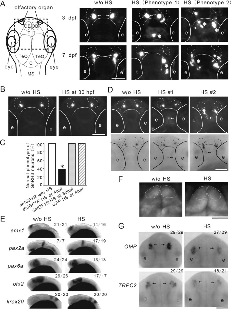
IGF signaling regulates the spatial organization of GnRH3 neurons; this action is developmental stage-dependent. A, Blockade of IGF signaling in early embryos results in abnormal distribution of GnRH3 neurons in the larval brain. Left, Diagram illustrating the orientation and anatomy of zebrafish larval brain. OB, olfactory bulb; T, telencephalon; TeO, tectum opticum; C, cerebellum; MS, medulla spinalis. Tg(hsp70:dnigf1r-GFP/GnRH3:EGFP) double-transgenic embryos were treated as described in Figure 2A and raised to 3 and 7 dpf. The location of GFP-positive GnRH3 neurons was visualized and representative images are shown (right). GnRH3 neurons at the two normal locations are indicated by arrows. Aberrant GnRH3 neurons are indicated by asterisks. Solid lines indicate the locations of eyes (e). Broken lines represent olfactory organs. Dashed line denotes the boundary between the olfactory bulb and telencephalon. B, Inhibition of IGF1R in advanced embryos has no effect. Tg(hsp70:dnigf1r-GFP/GnRH3:EGFP) double-transgenic embryos were subjected to 1 h HS treatment at 30 hpf. The embryos were raised to 3 dpf and the distribution of GnRH3 neurons was examined and shown. C, Quantitative results of A and B. n = 19∼41, *p < 0.001 compared with the no (w/o) HS group. D, The ectopic GFP-positive cells are indeed GnRH3 neurons. Larvae (3 dpf old) described in A were subjected to in situ hybridization analysis for GnRH3mRNA. Top, GFP view; bottom, in situ hybridization results. Aberrant GnRH3 neurons are indicated by arrows. Similar results were obtained in all eight embryos examined. E, Blockage of IGF signaling has no effect on global brain patterning. Tg(hsp70:dnigf1r-GFP) embryos were treated as described in A, raised to 32 hpf, and subjected to in situ hybridization analysis for the indicated marker genes. The frequency of embryos with the indicated expression patterns is shown in the upper right of each panel. Scale bar, 0.2 mm. F, Blockage of IGF signaling does not alter the morphology of olfactory bulbs. Tg (α1-tubulin1a: EGFP/hsp70:dnIGF1R-GFP) embryos are treated as described in A, raised to 3 dpf, and analyzed. Dashed line denotes the olfactory bulbs. G, Blockage of IGF signaling does not alter the location of specific sensory neurons. Tg(hsp70:dnigf1r-GFP) embryos were treated as described in A, raised to 32 hpf, and subjected to in situ hybridization analysis for the indicated marker genes. Arrows indicate locations of olfactory sensory neurons expressing OMP and TRPC2.
