Figure 5.
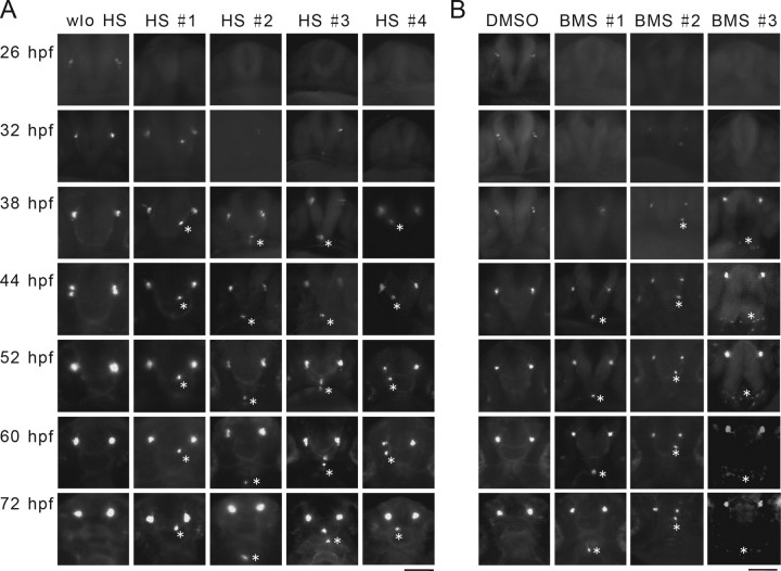
IGF signaling regulates the proper spatial distribution of newborn GnRH3 neurons. A, Time-lapse imaging of GnRH3 neurons in Tg(hsp70:dnigf1r-GFP/GnRH3:EGFP) double-transgenic fish. Embryos were treated as described in Figure 2A. The timing of emergence and spatial location of GnRH3 neurons were monitored in the same individuals at the indicated time points. Four HS-treated fish (#1–4) are shown. B, Time-lapse imaging of GnRH3 neurons in Tg(GnRH3:EGFP) fish. Embryos were treated with DMSO or BMS-754807 (BMS; 3 μm) from 4 to 32 hpf. The timing of emergence and spatial location of GnRH3 neurons were monitored in the same individuals at the indicated time points. Three treated fish (#1–3) are shown. Aberrant GnRH3 neurons are indicated by asterisks.

