
Figure 2.
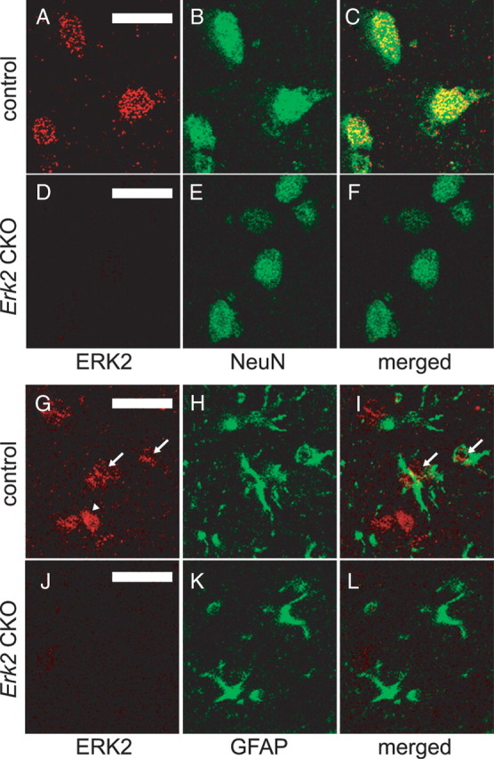
ERK2 protein is abrogated in neuronal and glial cells in Erk2 CKO mice. A–C, ERK2 is expressed in neuronal cells in control mice at 12 weeks of age. Double staining for ERK2 (A) and the postmitotic neuronal marker NeuN (B) in the neocortex show that ERK2 is expressed in neurons, as indicated by colocalization (C). D–F, ERK2 is abrogated in neuronal cells in Erk2 CKO mice. Double staining for ERK2 (D) and NeuN (E) with a merged image (F) show that ERK2 is not detectable in neuronal cells in Erk2 CKO mice. G–I, ERK2 is expressed weakly in astrocytes in control mice. Double staining for ERK2 (G) and the astrocyte marker GFAP (H) in the neocortex with a merged image (I) show partial colocalization of ERK2 and GFAP, indicating that ERK2 is expressed weakly in some astrocytes (arrows) although not in other astrocytes. Arrowhead indicates probable expression of ERK2 in neurons. J–L, ERK2 is abrogated in astrocytes in Erk2 CKO mice. Double staining for ERK2 (J) and GFAP (K) with a merged image (L) show that ERK2 is not detectable in astrocytes in Erk2 CKO mice. Scale bars, 20 μm.