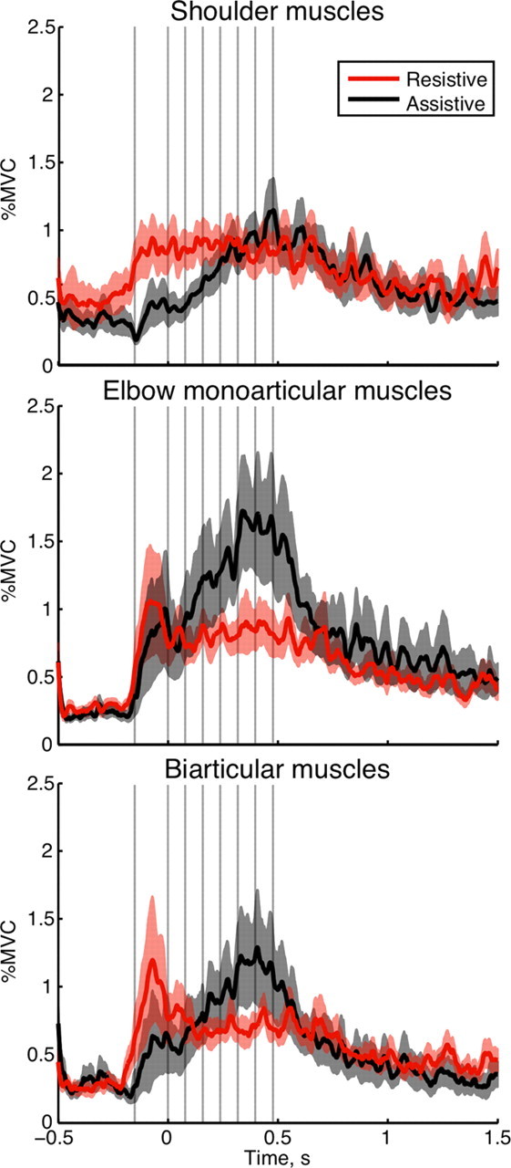
Figure 5.

Co-contraction between antagonistic muscles across subjects. Top plot, Posterior deltoid with pectoralis major; middle plot, brachioradialis with triceps lateral; bottom plot, biceps long with triceps long. The traces show mean co-contraction in time during the resistive (red) and assistive (black) conditions; the shaded areas outline SEM across subjects. The vertical gray lines indicate times of TMS across multiple trials; zero time is the moment of exiting the T0 target.
