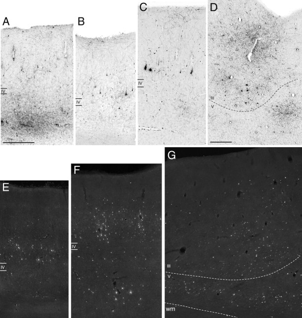
Figure 6.

Examples of laminar patterns of retrograde and anterograde labeling, observed after injections in intermediate area 12r, in areas 11 (A), TEa/m (B, E), SII (C, F), and F5a (D, G). A and B are from Case 44r FR, C and D from Case 44r LYD, and E–G from Case 43l FB. Scale bars: A, D, 500 μm (A also applies to B, C, E–G). wm, White matter.
