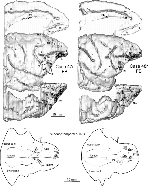
Figure 7.
Distribution of the retrograde labeling observed after injections in rostral area 12r in Cases 47r FB and 48r FB, shown in dorsolateral, medial, and bottom views of the 3D reconstructions of the injected hemispheres (top) and in 2D reconstructions of the STS (bottom). Conventions and abbreviations as in Figures 1 and 3.

