Figure 3.
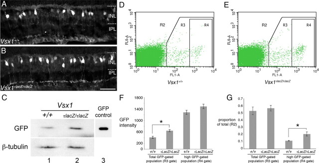
Upregulation of the GUS8.4GFP reporter transgene in Vsx1τLacZ/τLacZ mice. A, B, GUS8.4GFP reporter immunolabeling in the Vsx1+/+ (A) and Vsx1τLacZ/τLacZ (B) retina. IPL, Inner plexiform layer; INL, inner nuclear layer. C, Western blot showing GFP and control β-tubulin protein levels from total retinal lysates of Vsx1+/+ (column 1) and Vsx1τLacZ/τLacZ (column 2) mice. 293 cells transfected with a GFP-expressing plasmid were used as a positive control for GFP detection (column 3). D–G, Flow cytometry data were obtained from papain-dissociated retinas from Vsx1+/+ and Vsx1τLacZ/τLacZ mice carrying the GUS8.4GFP transgene. Representative examples showing the forward and side scatter of cell populations designated as R2 (i.e., all of the cells within the plot) are shown for Vsx1+/+ (D) and Vsx1τLacZ/τLacZ (E) mice. E, Relative mean fluorescence in the GFP-positive population indicated by R3 (i.e., all of the cells within the R3 box) was significantly higher in Vsx1τLacZ/τLacZ mice (Vsx1+/+, 411 ± 39; Vsx1τLacZ/τLacZ, 643 ± 36; Student's t test p < 0.05). Within the subpopulation of higher GFP-expressing cells (R4), mean fluorescence intensity was not significantly increased (F), indicating that highly fluorescing cells in Vsx1+/+ mice were not getting brighter in Vsx1τLacZ/τLacZ mice. Although the total number of GFP fluorescing cells (R3/R2) was unchanged in Vsx1tLacZ/tLacZ mice (G), a significant increase in the number of cells with high GFP fluorescence (R4 /R2) was observed (F, G, *p < 0.05 by Student's t test). Scale bar: (in B) A, B, 35 μm.

