
Figure 4.
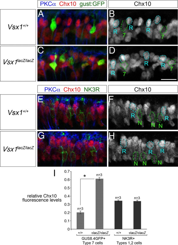
Upregulation of Chx10 in Type 7 bipolar cells in Vsx1τLacZ/τLacZ mice. A–H, Immunolabeling of adult retinal sections for Chx10, PKCα, and the GUS8.4GFP (gust:GFP) reporter (A–D) and Chx10, PKCα, and NK3R (E–H). Chx10 immunofluorescence in Type 7 bipolar cells is outlined in green and labeled “7” in B and D. Chx10 immunofluorescence in PKCα-positive rod bipolar cells is outlined in blue and labeled “R” in B, D, F, and H. NK3R-immunolabeled Type 1 and 2 OFF bipolar cells are outlined in green lines and labeled “N” in F and H. I, Chx10 immunofluorescence levels in Type 7 or Type 1/2 bipolar cells was normalized to Chx10 immunofluorescence levels in rod bipolar cells and compared in Vsx1+/+ and Vsx1τLacZ/τLacZ mice. In the Vsx1τLacZ/τLacZ retina, Chx10 immunofluorescence is upregulated in Type 7 bipolar cells to almost three times of that observed in the wild-type retina (0.610 ± 0.018 in Vsx1τLacZ/τLacZ compared to 0.202 ± 0.018 in Vsx1+/+, mean ± SE, n = 3). The level of Chx10 immunofluorescence does not change significantly in NK3R immunolabeled OFF bipolar cells in the Vsx1τLacZ/τLacZ versus the Vsx1+/+ retina (0.338 ± 0.011 in Vsx1τLacZ/τLacZ compared to 0.342 ± 0.010 in Vsx1+/+, mean ± SE, n = 3). The asterisk in I indicates a significant difference by Student's t test (*p < 0.01). Scale bar: (in D) A–H, 14 μm.