
Figure 5.
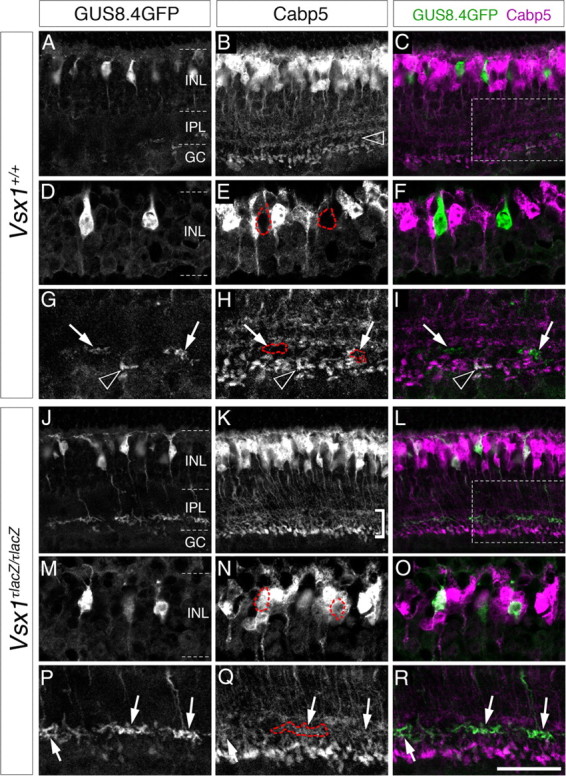
Cabp5 is ectopically expressed in Type 7 bipolar cells in Vsx1τLacZ/τLacZ mice. In the Vsx1+/+ retina, Calcium-binding protein 5, Cabp5, is expressed in a subset of bipolar cells (Type 3 OFF and Type 5 ON and rod bipolar cells) (Haverkamp et al., 2003) and is normally not detected in putative Type 7 bipolar cells distinguished by their high level of fluorescence (A–I). The absence of Cabp5 in Type 7 cells is highlighted by red dashed for GFP-expressing soma (E) and axon terminals (G–I, arrows). The black arrowhead in G–I points to the axon terminal of a GFP-expressing rod bipolar cell (distinguished by its position and shape) that is colabeled with Cabp5. In the Vsx1+/+ retina, a characteristic gap in Cabp5 immunolabeling is observed between the axon terminals of Type 5 and rod bipolar cells (B, open arrowhead); however this gap is no longer evident in the Vsx1τLacZ/τLacZ retina [bracketed region in K (Chow et al., 2004)]. Within this region of the inner plexiform layer, Cabp5 immunolabeling colabels with GFP in the axon terminals of Type 7 bipolar cells (J–L; arrows in P–R and red-outlined axon terminals in Q). Colocalization of Cabp5 immunolabeling with the GUS8.4GFP reporter in the Vsx1τLacZ/τLacZ retina was also observed in the cell bodies of putative Type 7 cells (M–O, outlined in red in N). The boxed regions in C and L are shown at higher magnification in (G–I) and (P–R), respectively. IPL, Inner plexiform layer; INL, inner nuclear layer; GC, ganglion cell layer. Scale bar: (in R) A–C, J, K, 40 μm; D–G, M–R, 20 μm.