Figure 8.
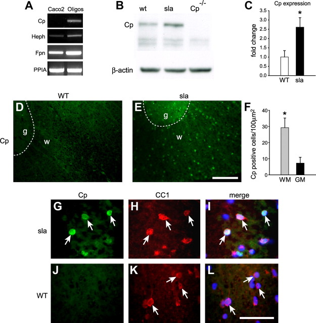
Expression of Cp is upregulated in white matter oligodendrocytes in sla mice. A, RT-PCR analysis shows that cultured oligodendrocytes express mRNA for Heph, Fpn, as well as Cp. In contrast, oligodendrocytes do not express Cp protein (data not shown). RNA isolated from the intestinal cell line Caco2 is included as a positive control for Heph and Fpn and a negative control for Cp. PPIA levels demonstrate that equal amounts of RNA were used. B, Western blot shows that Cp expression is increased in the spinal cord of sla mice compared with wild-type (WT) mice. Spinal cord protein extract of Cp KO (Cp−/−) mice was used as a negative control. The blot was reprobed for β-actin to ensure equal protein loading. C, Quantification of the Western blot data shows a significant increase of Cp expression in the spinal cord of sla mice. Results are shown as mean ± SEM (n = 3; *p ≤ 0.05, Student's t test). D, E, In situ hybridization for Cp on spinal cord cross sections shows upregulation of Cp expression in the white matter of sla mice (E) compared with wild-type mice (D). The dashed lines outline the boundary between gray (g) and white (w) matter. F, Quantification of the number of Cp-positive cells in the gray and white matter of sla spinal cord shows that Cp induction is markedly greater in the white matter compared with the gray matter. Results are shown as mean ± SEM. n = 3; *p ≤ 0.05, Student's t test. G–I, Higher magnification of the sla spinal cord white matter showing in situ hybridization for Cp (G) combined with immunofluorescence labeling with anti-CC1 for oligodendrocytes (H), indicating that Cp expression is upregulated in white matter oligodendrocytes (arrows). J–L, In contrast, there is no Cp expression (J) in CC1-positive oligodendrocytes (K) in wild-type (WT) mice. I and L show merged images. Scale bars: E, 100 μm; L, 50 μm. WM, White matter; GM, gray matter; PPIA, peptidylprolyl isomerase A.

