
Figure 1.
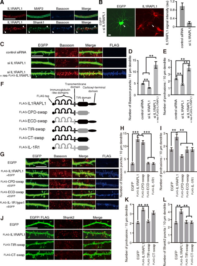
Effects of IL1RAPL1 on synapse formation. A, Immunostaining of cultured cortical neurons with antibodies against IL1RAPL1, MAP2, and Bassoon (top) and with those against IL1RAPL1, Shank1, and Bassoon (bottom). B, Efficacy of siRNAs against Il1rapl1 in cultured cortical neurons. Cortical neurons were transfected with siRNAs against Il1rapl1 together with an expression vector for EGFP and were immunostained for IL1RAPL1. siRNAs against Il1rapl1 reduced IL1RAPL1 staining signal intensity by 80% compared with control siRNA. C, Reduction of numbers of Bassoon puncta and dendritic protrusions of cultured cortical neurons by siRNAs against Il1rapl1 and rescue by res-FLAG-IL1RAPL1. D, E, Effects of siRNA treatments on the numbers of Bassoon puncta (D) and dendritic protrusions (E) along dendrites of cortical neurons. n = 52, 52, and 20 neurons for control siRNA, siRNA against Il1rapl1 mRNA, and siRNA against Il1rapl1 mRNA with res-FLAG-IL1RAPL1, respectively. F, Schematic structures of FLAG-IL1RAPL1, FLAG-IL-1R1, and their swap mutants. G, Effects of IL1RAPL1/IL-1R1 swap mutants on dendritic protrusions and Bassoon puncta in cultured cortical neurons. H, I, Numbers of Bassoon puncta (H) and dendritic protrusions (I) along dendrites of cortical neurons transfected with EGFP, FLAG-IL1RAPL1, FLAG-CPD-swap, FLAG-ECD-swap, and FLAG-IL-1R1 (n = 28–40 neurons). J, Effects of TIR and C-terminal domain-swap mutants on dendritic protrusions and Shank2 puncta in cultured cortical neurons. K, L, Numbers of dendritic protrusions (K) and Shank2 puncta (L) along dendrites of cortical neurons transfected with EGFP, FLAG-IL1RAPL1, FLAG-TIR-swap, and FLAG-CT-swap (n = 19–30 neurons). All values represent mean ± SEM. **p < 0.01, ***p < 0.001, Tukey's test. Scale bars, 10 μm.