
Figure 2.
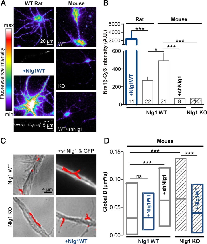
Downregulating Nlg1 expression increases AMPAR membrane diffusion. A, Live labeling of endogenous surface Nlg1 using soluble Nrx1β-Fc in 1- week-old rat or mouse neurons, upon Nlg1 overexpression or downregulation. The clustered appearance of overexpressed Nlg1 was comparable to that of endogenous Nlg1 (insets). Nlg1 cluster density was 0.24 ± 0.03/μm2 for transfected cells versus 0.08 ± 0.01/μm2 for nontransfected cells, and cluster size was 0.51 ± 0.02 μm2 versus 0.24 ± 0.02 μm2, respectively (n = 7 neurons). B, Quantification of the Cy3-conjugated anti-Fc labeling in arbitrary fluorescence units. The number of cells examined in each condition is indicated in the columns (2 independent experiments). C, AMPAR diffusion was measured using anti-GluA2-conjugated Qdots in primary 9–10 DIV neurons obtained from either WT or Nlg1 KO mouse pups. In some experiments, KO cultures were transfected with Nlg1WT, and WT cultures with Nlg1WT or shRNA against Nlg1. D, Diffusion coefficients were computed for 150–2400 trajectories from 6 independent experiments. Statistical P-values: *p < 0.05; ***p < 0.001.