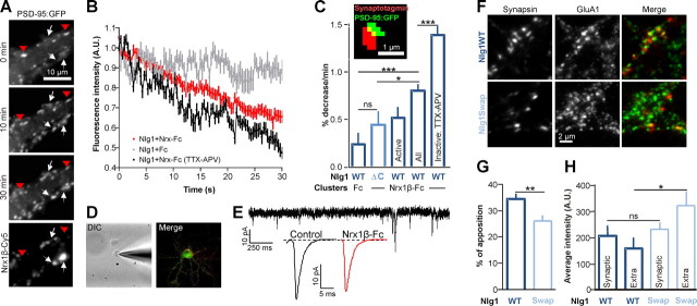
Figure 8.
Recruitment of PSD-95 and AMPARs at novel Nrx/Nlg adhesions competes with preexisting synapses. A–C, Rat hippocampal neurons (9 DIV) expressing either Nlg1WT or Nlg1ΔC, together with PSD-95:GFP were incubated with purified Nrx1β-Fc (or human Fc as a control) cross-linked by Cy5 labeled anti-Fc antibodies. In some experiments, neurons were treated throughout the recordings with TTX/APV, to inhibit synaptic activity. The distribution of PSD-95:GFP was monitored in living cells during a 30 min. A, Representative time course showing the progressive disappearance of PSD-95:GFP from preexisting clusters (red arrowheads), concomitant to the formation of new clusters (white arrows) upon Nrx1β-Fc addition. B, Quantification of the fluorescence decay of preexisting PSD-95:GFP clusters, for 3 conditions (control Fc, Nrx1β-Fc untreated, and Nrx1β-Fc treated with TTX/APV). C, Depletion rates were quantified by fitting a linear relationship through the fluorescence decay for each cluster (mean ± SEM of 27–92 clusters from 6–16 cells). In some experiments, active synapses were labeled using a synaptotagmin antibody uptake, and PSD-95:GFP clusters apposed to synaptotagmin puncta (inset) were pooled as a separate group. D, E, Neurons expressing Nlg1WT and PSD-95:GFP either without treatment or after a 1 h incubation with cross-linked Nrx1β-Fc, were subjected to patch-clamp recording, in the presence of TTX and APV to isolate AMPA mEPSCs. D, Images of a patched neuron in DIC and in fluorescence (PSD-95:GFP in green, Nrx1β-Fc Cy5 clusters in red). E, Representative time sequence and average AMPA mEPSCs traces. F, Double staining of endogenous AMPAR with anti-GluA1 antibodies (green), and presynapses with anti-synapsin (red), in 10 DIV neurons expressing either Nlg1WT or Nlg1Swap. Transfected cells were detected by coexpression of GFP (data not shown). G, Analysis of the fraction of GluA1 clusters apposed to synapsin spots, either upon Nlg1WT or Nlg1Swap expression. (n = 20 and 15 cells, respectively, from 2 independent experiments). H, Neurons (10 DIV) expressing either Nlg1WT or Nlg1Swap and PSD-95:GFP, were live-labeled for endogenous AMPAR with anti-GluA1 antibody, and with anti-synapsin to stain presynapses (data not shown). The graph represents the intensity of GluA1 in puncta apposed to presynapses or colocalizing with PSD-95:GFP clusters nonapposed to synapsin (n = 12 cells for each condition from 2 independent experiments). Statistical P-values: *p < 0.05, **p < 0.01, ***p < 0.001.

