
Figure 3.
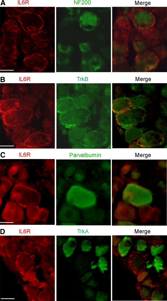
IL-6R is expressed in a subset of myelinated sensory neurons. A, Apotome images of transversal slices of axotomized DRG double stained with anti-IL-6R (red) and anti-NF-200 (green) antibodies. The merge image shows colocalization of IL-6R with NF-200-positive sensory neurons. B, Double immunochemistry using anti-TrkB antibody (green) shows IL-6R colocalization with TrkB-positive neurons (merge). C, Double immunochemistry using anti-Parvalbumin antibody (green) shows IL-6R colocalization with parvalbumin-positive neurons (merge). D, Double immunochemistry using anti-TrkA antibody (green) demonstrates no IL6-R colocalization with TrkA-positive neurons (merge). Scale bar, 30 μm.