
Figure 8.
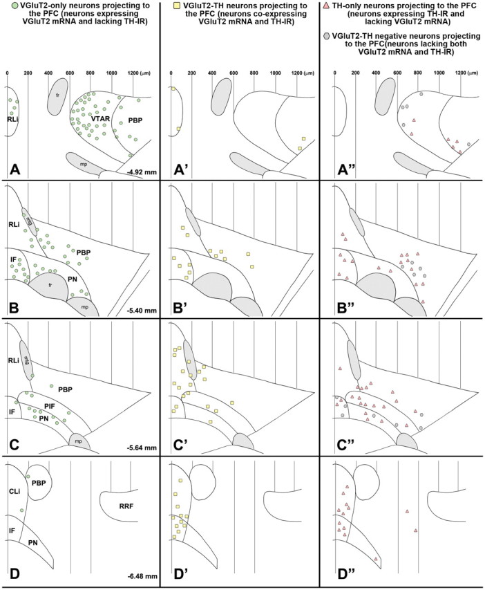
Summary diagram of the rostrocaudal and mediolateral distribution of four different phenotypes of mesocortical neurons. A–D, Mesocortical VGluT2-only neurons (FG-labeled neurons expressing VGluT2 mRNA but lacking TH immunoreactivity) with a decreasing rostrocaudal gradient of distribution. A′–D′, Mesocortical VGluT2-TH neurons (FG-labeled neurons coexpressing VGluT2 mRNA and TH immunoreactivity) are concentrated in the caudal aspects of both the RLi and the IF. A″–D″, Mesocortical TH-only neurons (FG-labeled neurons lacking expression of VGluT2 mRNA but containing TH immunoreactivity) are intermingled with mesocortical VGluT2-TH-negative neurons (FG-labeled neurons lacking both VGluT2 mRNA and TH immunoreactivity). The symbols in each panel represent the pool of FG-labeled neurons found in four brain sections, with each section selected from a different rat. mtg, Mammillotegmental tract; fr, fasciculus retroflexus; mp, mammillary peduncle; VTAR, ventral tegmental area rostral; RRF, retrorubral field.