Figure 1.

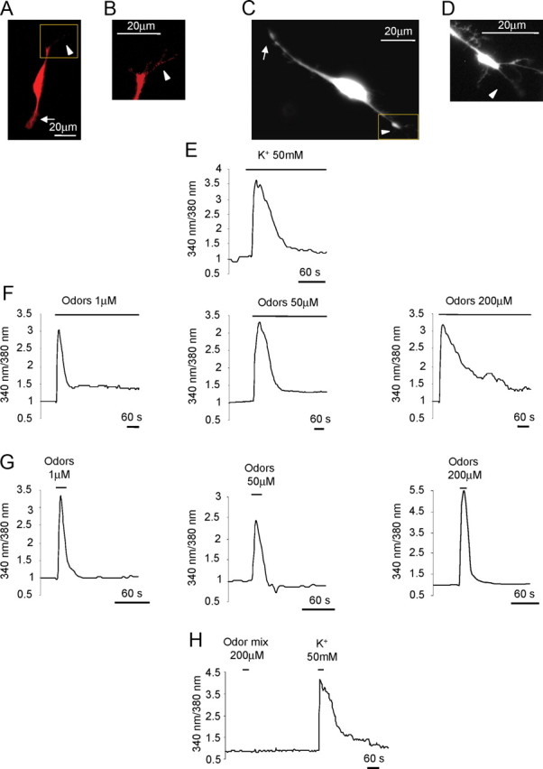
Ca2+ dynamics in OSN in culture. A, Example of an OSN immunopositive for OMP. B, Higher magnification of the cilia indicated in the square in A. C, Example of an OSN loaded with fura-2. D, Higher magnification of the cilia emanating from the knob, indicated in the square in C. Arrows, Axon terminus-growth cone; arrowheads, cilia-dendrite. Scale bar, 20 μm. E–H, Normalized fluorescence ratio changes (340/380 nm) in OSN loaded with fura-2 and challenged with KCl (50 mm), bath applied (E); odor mixture at 1, 50, 200 μm, bath applied (F); odor mixture at 1, 50, 200 μm, bath applied for 4–10 s (G); example of a nonresponsive neuron to the odor mixture (200 μm, bath applied for 4–10 s), but responsive to KCl (50 mm) bath applied for 4–10 s (H). Primary culture of OSNs were used in all experiments.
