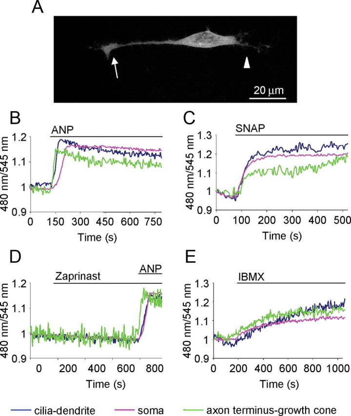
Figure 2.

cGMP dynamics in OSN upon pharmacological stimuli. A, Example of an OSN transfected with the sensor for cGMP. The fluorescence is distributed throughout the cytoplasm with the exclusion of the nucleus. Arrow, Axon terminus-growth cone; arrowhead, cilia-dendrite. Scale bar, 20 μm. B–E, Normalized kinetics of fluorescence emission intensities (480/545 nm) recorded in cilia-dendrite, soma, and axon terminus-growth cone in OSNs transfected with the PKG-based sensor for cGMP and challenged with different stimuli, all bath applied via an application pipette positioned far away (∼3 mm) from the cell, to obtain a homogeneous distribution of the stimuli in the bath. B–E, ANP (1 μm) activator of mGC (B); SNAP (300 μm) NO donor, which activates sGC (C); zaprinast (250 μm), inhibitor of the cGMP-specific PDE-5 and subsequently with ANP (1 μm) (D); and IBMX (250 μm), nonselective inhibitor of PDEs (E). Neurons challenged only by Ringer's solution with or without the solvent (negative controls), did not present changes in fluorescence ratio, here and in all the subsequent treatments (data not shown). The regions of interest were drawn on the distal portion of the axon (axon terminus-growth cone), on the soma, and on the distal part of the dendrite (cilia-dendrite). Other conditions as in Materials and Methods. Blue line, CFP/YFP in cilia-dendrite; pink line, CFP/YFP in soma; green line, CFP/YFP in axon terminus-growth cone. Primary cultures of OSNs were used in all experiments.
