Figure 5.

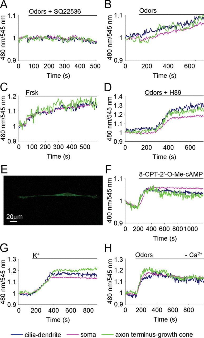
Molecular mechanism underpinning cGMP rise. Conditions as in Figure 2. A, B, Examples of the spatiotemporal dynamics of cGMP in the same OSN treated with odors (200 μm) in presence of AC inhibitor SQ22536 (30 μm) (A) and with odors only (200 μm) after washing away the inhibitor SQ22356 (B). C, D, OSNs treated with Frsk (25 μm), AC activator (C), and odors (200 μm) in the presence of the PKA inhibitor H89 (10 μm) (D). E, Example of an OSN immunopositive for Epac1. The immunofluorescence is present in the entire neuron. Scale bar, 20 μm. F–H, Examples of cGMP dynamics in OSNs treated with Epac activator 8-CPT-2′-O-Me-cAMP (30 μm) (F), KCl (50 mm) (G), and odors (200 μm), in a Ca2+-free Ringer's solution (H). Stimuli were all bath applied. Blue line, Cilia-dendrite; pink line, soma; green line, axon terminus-growth cone. Primary cultures of OSNs were used in all experiments.
