Figure 1.
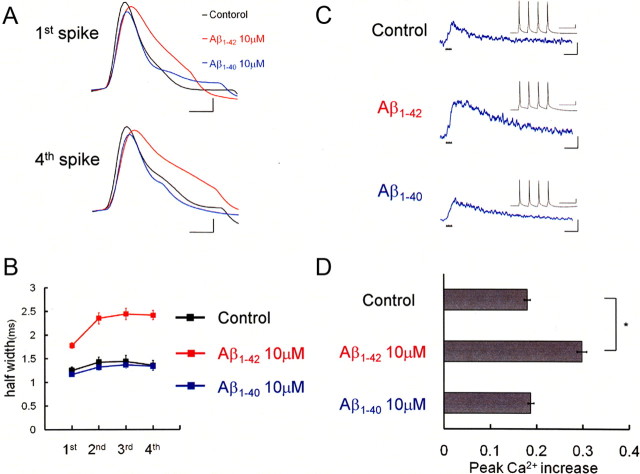
Intracellular infusion of Aβ1-42 broadens spike width and augmented Ca2+ influx in rat neocortical pyramidal neurons. A, Action potentials evoked in neurons injected with Aβ. The first and fourth action potentials in spike trains are shown. Recordings taken from the control neuron (black), an Aβ1-42-injected neuron (red), and an Aβ1-40-injected neuron (blue) are superimposed to clarify the spike broadening in Aβ1-42-injected neurons. Calibration: 1 ms, 20 mV. B, Average spike half-width in four-spike trains, showing that injection of Aβ1-42 (10 μm; red square; n = 6), but not that of Aβ1-40 (10 μm; blue square; n = 6), broadened spike width compared with control neurons (black square; n = 6). C, Ca2+ increases induced by four-spike trains (inset) in control neurons and in neurons injected with 10 μm Aβ1-42 or 10 μm Aβ1-40. Calibration: 500 ms, −0.1ΔF380/F360. Inset, Specimen recording of a spike train at 18 Hz that was used for the shown Ca2+ measurements. The timing of each spike is shown by a small black triangle below the trace. Note the difference in time scale. Calibration: 100 ms, 20 mV. D, Summary diagram demonstrating average Ca2+ increases. Injection of Aβ1-42 enhanced spike-induced Ca2+ increases. *p < 0.0001.

