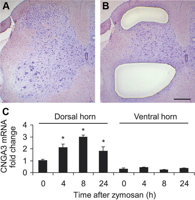
Figure 2.
CNGA3 expression in the ipsilateral dorsal and ventral horn of the spinal cord after intraplantar zymosan injection. A–C, Spinal cord sections were stained with cresyl violet (A), and tissues of the superficial dorsal horn (laminae I–III) and ventral horn (laminae VII–IX) were collected using laser microdissection (B). Total RNA was isolated from collected tissues and subjected to real-time RT-PCR analysis (C). The quantity of CNGA3 mRNA relative to GAPDH mRNA was calculated by the 2−ΔΔCT method. n = 3 per group. Note that in naive animals, more CNGA3 mRNA was detected in the superficial dorsal horn compared with the ventral horn, and that zymosan-induced CNGA3 upregulation occurs only in the superficial dorsal horn. Scale bar, 250 μm.

