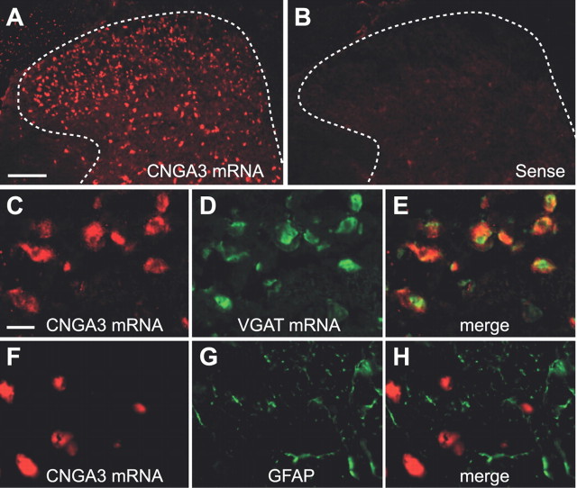
Figure 3.
Cellular localization of CNGA3 in the spinal cord. A, In situ hybridization using an antisense probe for CNGA3 reveals high levels of CNGA3 mRNA expression within the superficial dorsal horn. B, Specificity of the CNGA3 antisense probe was confirmed by incubating with the respective sense probe directed against the complementary mRNA sequence. Dotted lines delineate gray matter. C–E, Double in situ hybridization of CNGA3 mRNA and VGAT mRNA. F–H, Double labeling by in situ hybridization of CNGA3 mRNA and immunohistochemistry for GFAP. CNGA3 mRNA was detected using HNPP (A, C) or TSA cyanine 3 (F) and appears in red. Markers were detected using TSA fluorescein (D) or Alexa Fluor 488 (G) and appear in green. Pictures C–H were taken from lamina II. Data indicate that CNGA3 is mainly expressed in inhibitory neurons of the superficial dorsal horn. Scale bars: A, 100 μm; C, 10 μm.

