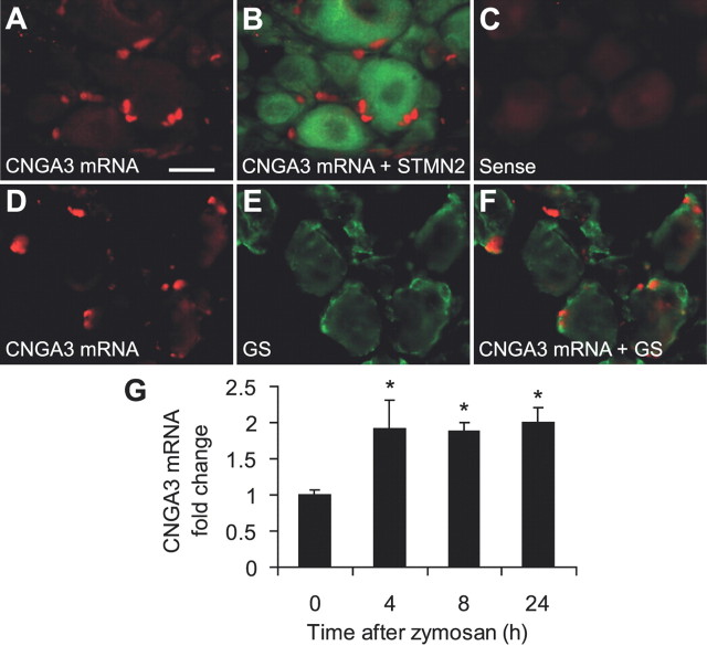
Figure 4.
Cellular localization of CNGA3 in dorsal root ganglia. A, B, In situ hybridization experiments detected CNGA3 mRNA (red) in DRGs (A), and double labeling with immunohistochemistry for the neuronal marker stathmin-like 2 (STMN2; green) indicates that CNGA3 mRNA is not expressed in DRG neurons (B). C, Specificity of the CNGA3 antisense probe was confirmed by incubating with the respective sense probe directed against the complementary mRNA sequence. D–F, Double labeling by in situ hybridization of CNGA3 mRNA and immunohistochemistry for the satellite glial cell marker glutamine synthetase (GS; green) suggests CNGA3 to be mainly expressed in satellite glial cells. G, Real-time RT-PCR analyses demonstrating that CNGA3 mRNA was significantly upregulated in DRGs 4–24 h after intraplantar zymosan injection. The quantity of CNGA3 mRNA relative to GAPDH mRNA was calculated by the 2−ΔΔCT method. n = 4 per group. Data are presented as mean ± SEM. *p < 0.05. Scale bar, 20 μm.

