
Figure 7.
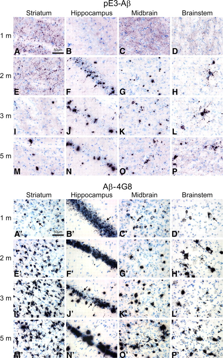
Age- and region-dependent accumulation of Aβ and pE3–Aβ in HOM TBA2.1 mice. For a simultaneous front-to-back representation of 25 brains, high-sensitivity immunohistochemistry was performed on MultiBrain sections. Two-dimensional analysis of pE3–Aβ (A–P) and Aβ (A′–P′) immunoreactivity in TBA2.1 mice reveals differential distribution patterns in different brain regions of HOM TBA2.1 mice. Aβ reactivity using the 4G8 anti-Aβ antibody is found both intracellularly in intact cells (e.g., A′–C′, arrows) and extracellularly in evidently disintegrated cells (e.g., J′, arrows). Intracellular Aβ deposits appear to be originating from small, inclusion-like structures (D′, arrow) and seem to be deposited extracellularly after disintegration of the neuron (N′). In contrast, using both a newly generated anti-pE3–Aβ antibody and a commercially available antibody, highest reactivity of pE3–Aβ is detected extracellularly after neuronal disintegration (A–P).