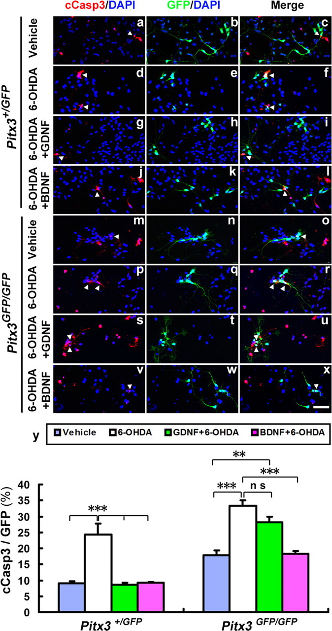
Figure 10.

GDNF does not protect against 6-OHDA-induced apoptotic cell death of Pitx3-null mutant mdDA neurons. a–x, Primary VM cultures derived from E11.5 Pitx3+/GFP (a–l) and Pitx3GFP/GFP (m–x) embryos were treated after 3 DIV with vehicle (0.1% BSA) (a–f, m–r), 20 ng/ml GDNF (g–i, s–u), or 20 ng/ml BDNF (j–l, v–x); after 2 h, cells were incubated with 10 μm 6-OHDA (d–l, p–x) or vehicle (PBS; a–c, m–o) for 24 h and immunostained for cCasp3 (red in a, d, g, j, m, p, s, and v) and GFP (green in b, e, h, k, n, q, t, and w); blue, DAPI stain; merged images in c, f, i, l, o, r, u, and x. y, Quantification of the relative amount of apoptotic (cCasp3+) mdDA neurons (GFP+) in these eight experimental groups revealed that both GDNF and BDNF prevent the 6-OHDA-induced cell death of Pitx3+/GFP mdDA neurons, but only BDNF (and not GDNF) prevents the 6-OHDA-induced cell death of Pitx3GFP/GFP mdDA neurons [cCasp3+/GFP+ cells (white arrowheads): Pitx3+/GFP vehicle-treated, 9.11 ± 0.55%; Pitx3+/GFP 6-OHDA-treated, 24.29 ± 3.49%; Pitx3+/GFP 6-OHDA + GDNF-treated, 8.70 ± 0.55%; Pitx3+/GFP 6-OHDA + BDNF-treated, 9.25 ± 0.11%; mean ± SEM, n = 3; Pitx3GFP/GFP vehicle-treated, 17.92 ± 1.46%; Pitx3GFP/GFP 6-OHDA-treated, 33.36 ± 1.74%; Pitx3GFP/GFP 6-OHDA + GDNF-treated, 28.11 ± 1.86%; Pitx3GFP/GFP 6-OHDA + BDNF-treated, 18.35 ± 0.78%, mean ± SEM; n = 3]. One-way ANOVA was used for statistical analysis of treatment effects. **p < 0.005; ***p < 0.001. Data were derived from three independent experiments. Scale bar: (in x), 50 μm.
