
Figure 5.
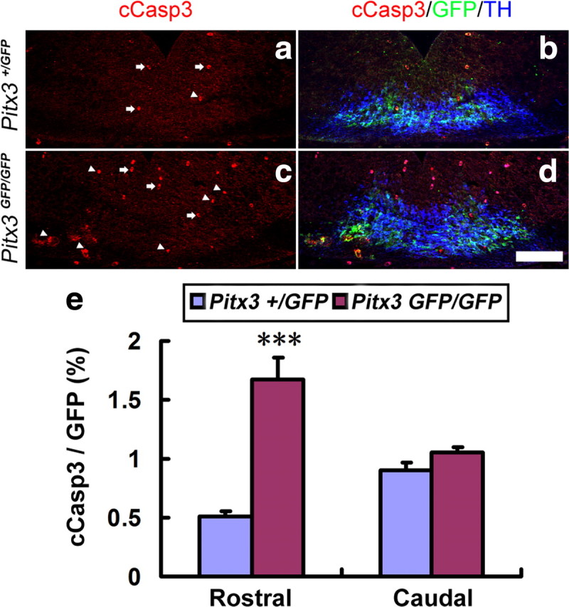
The loss of BDNF expression correlates with an increased apoptotic cell death of mdDA neurons in the E12.5 Pitx3GFP/GFP rostral VM. a–d, Triple immunostaining for cCasp3, GFP, and TH on representative midbrain coronal sections of Pitx3+/GFP (a, b) and Pitx3GFP/GFP (c, d) embryos at E12.5 revealed an increase of apoptotic Pitx3+ mdDA neurons [cCasp3+ (red) and GFP+ (green) double-labeled cells, white arrowheads) in the mutant VM. White arrows point at erythrocytes that were stained unspecifically for cCasp3. e, Quantification of cCasp3+/GFP+ double-labeled cells relative to the total amount of GFP+ (Pitx3+) cells in these sections showed a significant increase of apoptotic mdDA neurons in the rostral (cCasp3+/GFP+ cells: E12.5 Pitx3+/GFP, 0.506 ± 0.047%; E12.5 Pitx3GFP/GFP, 1.676 ± 0.185%; n = 3, mean ± SEM; ***p < 0.001 in the independent samples t test), but not caudal (cCasp3+/GFP+ cells: E12.5 Pitx3+/GFP, 0.902 ± 0.068%; Pitx3GFP/GFP, 1.056 ± 0.042%, n = 3, mean ± SEM), VM of the Pitx3GFP/GFP embryos. Scale bar: (in d), 100 μm.