
Figure 6.
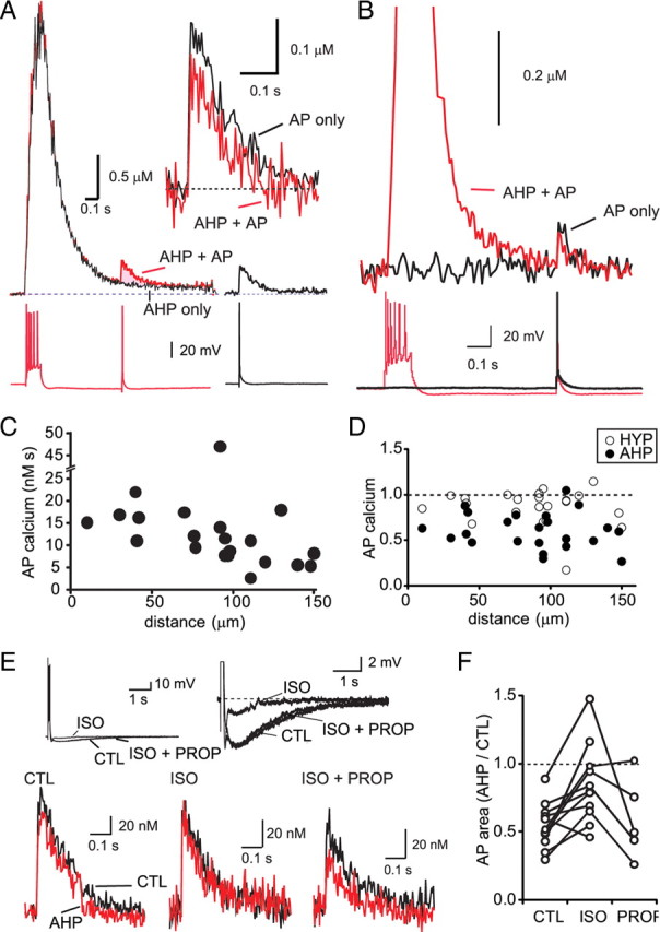
The sAHP reduces the dendritic AP-evoked calcium response. A, The AP-evoked calcium response is smaller during the sAHP. Top traces on the left show the calcium response in the dendrite (92 μm from the soma) in response to an AP train (black trace; AHP only) and to an AP train followed by a single AP evoked 500 ms after the train (red trace; AHP + AP). The calcium response evoked by the AP trains (AHP only) was subtracted from the paired response (AHP + AP) to determine the calcium rise evoked by the AP during the AHP (shaded area). The voltage response at the soma is shown below the calcium response. Calcium rise evoked by a single AP under control conditions is shown on the right. Inset (top right) shows the AP-evoked calcium rise overlaid with the AP-evoked calcium rise during the sAHP. B, An example of a dendritic calcium response (140 μm from the soma) in which there is little residual calcium from the AP train when the AP is evoked. In this instance, the peak response evoked by the control AP (black trace) is greater than the AP-evoked calcium rise during the sAHP (red trace). C, The AP-evoked calcium rise decreases with distance from the soma. The integrated area of the AP-evoked calcium rise in the dendrite is plotted against its distance from the soma. D, The AP-evoked dendritic calcium rise is attenuated during the sAHP but not during a hyperpolarizing somatic current injection that mimics the somatic hyperpolarization during the sAHP. The integrated area of the AP-evoked calcium rise during the sAHP (filled circles) and during a hyperpolarizing current injection (open circles) is plotted relative to control at various distances from the soma. E, F, Application of the β-adrenergic agonist isoprenaline reduces the sAHP and the attenuation of the AP-evoked calcium rise that follows the AP train. The somatic membrane potential is shown in response to a depolarizing current injection (100 ms) that evoked an AP train under control conditions (CTL) and after sequential application of isoprenaline (ISO) and the β-adrenergic receptor antagonist propranolol (PROP). The sAHP is shown on an expanded scale (right traces). The AP-evoked dendritic calcium rise before (blue) and during (red) the sAHP is shown under control conditions (bottom left traces), in the presence of isoprenaline (bottom middle traces), and in the presence of isoprenaline and propranolol (bottom right traces). F, Summary data showing the relative area of the single AP-evoked calcium rise during the sAHP under control conditions (CTL), after application of isoprenaline (ISO), and after addition of propranolol (PROP).