
Figure 1.
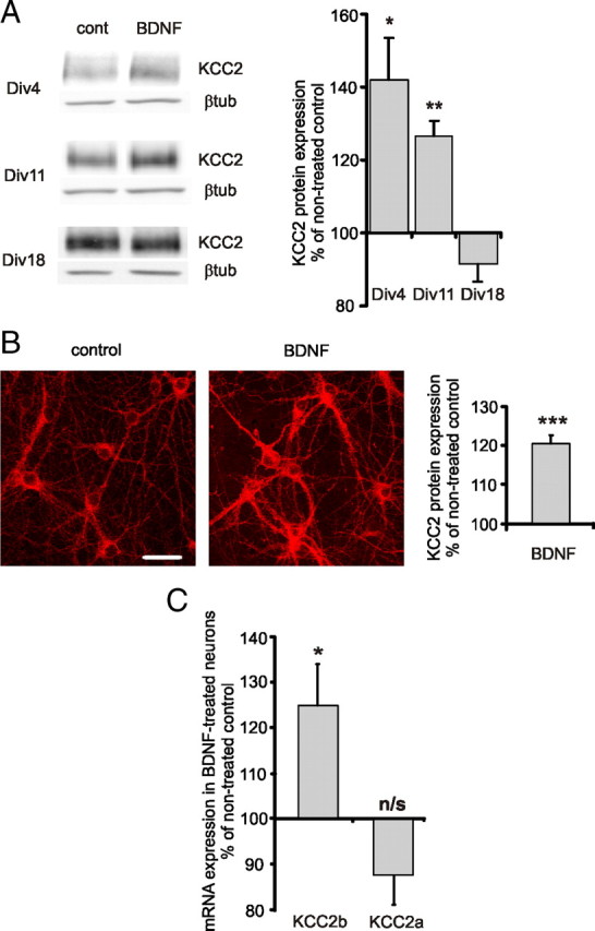
BDNF increases KCC2 expression in immature hippocampal cultures. A, Representative Western blot analysis and quantification of KCC2 expression in dissociated hippocampal cultures treated with BDNF (50 ng/ml) (n = 3–7). Dissociated cultures were treated with BDNF at DIV 1, DIV 8, and DIV 15, and analyzed 3 d after the treatment. Data are normalized with respect to the average in nontreated controls of corresponding age. *p < 0.05, **p < 0.01, one-sample t test. Error bars represent SEM. βtub, β-tubulin. B, Representative immunofluorescent staining and quantification of KCC2 expression in dissociated hippocampal cultures treated with BDNF (50 ng/ml) (n = 119–125). Neurons were treated with BDNF at DIV 6–8 and analyzed 24 h later. Data are normalized to the average in nontreated controls. ***p < 0.001, one-sample t test. Error bars represent SEM. Scale bar, 40 μm. C, KCC2b and KCC2a mRNA levels in DIV 6–8 cultured neurons 1–2 h after BDNF (50 ng/ml) application as detected by real-time PCR (n = 10). Values of corresponding nontreated controls were set to 100%. *p < 0.05, not significant (n/s); p > 0.05, one-sample t test. Error bars represent SEM.