
Figure 2.
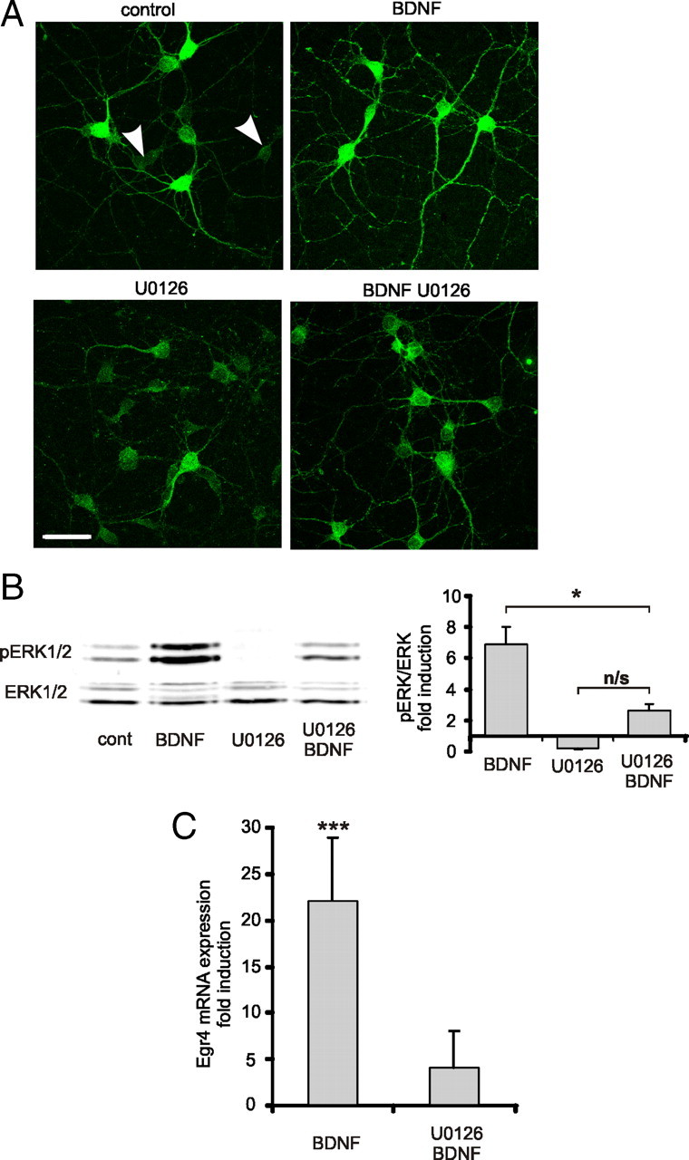
BDNF-induced Egr4 expression is dependent on ERK1/2 phosphorylation. A, Representative immunofluorescent staining of phosphorylated ERK1/2 (pERK1/2) in dissociated DIV 5–6 hippocampal neurons treated for 5 min with BDNF (50 ng/ml). Some of the cultures were pretreated with MEK inhibitor U0126 (20 μm) 30 min before BDNF treatment. Note that in nontreated control cultures there are neurons with very low levels of pERK1/2 (white arrowheads). In BDNF-treated cultures, all neurons exhibit high levels of pERK1/2 immunostaining intensity. Scale bar, 50 μm. B, Representative Western blot analysis and quantification of BDNF-induced ERK1/2 phosphorylation (n = 4). Cell lysates were collected 5 min after BDNF (50 ng/ml) application. Some cultures were pretreated with MEK inhibitor U0126 (20 μm) 30 min before BDNF application (n = 3). Data are normalized to the density of nonphosphorylated ERK1/2 bands and to a value in nontreated controls. *p < 0.05, not significant (n/s); p > 0.05; one-way ANOVA with subsequent Bonferroni's multiple-comparison test. Error bars represent SEM. C, Egr4 mRNA levels in dissociated DIV 5–6 hippocampal neurons 1–2 h after BDNF (50 ng/ml) application as detected by real-time PCR (n = 12). In some cases, cultures were pretreated with MEK inhibitor U0126 (20 μm) 30 min before BDNF application (n = 4). Nontreated control value set to 1. ***p < 0.001, one-sample t test. Error bars represent SEM.