
Figure 7.
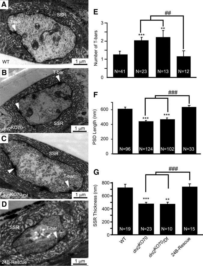
Ultrastructural analysis of type I synaptic bouton in dnl2 mutants. A–D, Representative transmission electron microscope micrographs showing low-magnification views of synaptic boutons, the presynaptic active zones (arrowheads), and postsynaptic SSR. E, Morphometric analysis of reconstructed synaptic boutons showed a significant increase in the number of T-bars per bouton in dnl2 mutants that was rescued by muscle specific expression of a UAS–dnl2 cDNA with 24B–Gal4. F, The length of PSDs beneath presynaptic T-bars was reduced in dnl2 mutants. This was also rescued by muscle expression of UAS–dnl2 with 24B–Gal4. G, The SSR was thinner in dnl2 mutants compared with WT controls or 24B–rescue flies. All images and analyses were derived from type Ib boutons on muscles 6/7. **p < 0.01 versus WT, ***p < 0.001 versus WT, ## p < 0.01 versus dnl2KO70, ### p < 0.001 versus dnl2KO70, Mann–Whitney test.