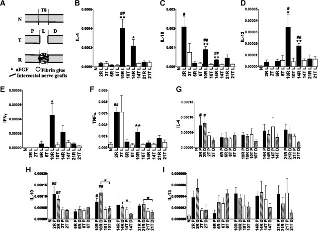
Figure 1.
The expression of cytokine IL-4, IL-10, and IL-13 is upregulated in the graft areas of repaired spinal cords. A, Group T, Normal rats (N) whose spinal cords were completely transected at T8 and for whom 5 mm of spinal cord tissue was removed. Group R, Spinal cord-transected rats that received a repair strategy of nerve grafts combined with aFGF in fibrin glue. P, Proximal stump; L, graft area of group R and lesion site of group T; D, distal stump. B–F, Q-PCR results revealed that the expression of TNFα (C), IL-4 (D), IL-10 (E), and IL-13 (F) was transiently induced 2 d after injury. In contrast, the graft areas of repaired spinal cords sustained the expression of these cytokines and IFN-γ (B). From 6 to 14 d, significantly higher IL-10 and IL-13 levels were found in repaired spinal cords compared with normal spinal cords, and the levels of IL-4, IL-10, and IL-13 increased in the graft areas of repaired spinal cords compared with injured spinal cords. #p < 0.05, ##p < 0.01 for group R or T compared with the normal group by Student's t test. *p < 0.05, **p < 0.01 for group R compared with group T by Student's t test. Determinations are means ± SEM from four to five experiments. G–I, IL-4 and IL-10 expression was increased in the stumps of 2 d repaired spinal cords compared with normal spinal cords. IL-10 expression was higher in the distal stumps of repaired spinal cords at 10–21 d than in transected spinal cords, but IL-4 and IL-13 levels were not increased in the stumps of repaired spinal cords compared with transected spinal cords. The levels in stumps were one-eighth of the levels in the graft areas at 10 d. *p < 0.05 between the indicated groups by Student's t test. Determinations are means ± SEM from four experiments.

