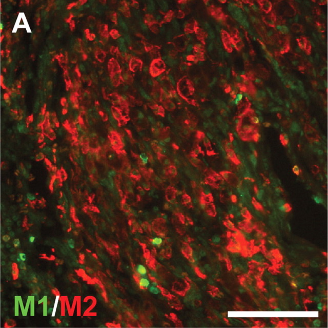
Figure 2.
M2 macrophages dominate within the graft areas of 14 d repaired spinal cords. Sections of 14 d repaired spinal cords were stained for CD86 (M1) and mannose receptor (CD206, M2). A large number of M2 macrophages were found in the grafted areas. Scale bar, 100 μm. Quantitation of macrophages expressing M1 and M2 markers in the grafted areas revealed that there were 74% M2 macrophages in the grafted areas of 14 d repaired spinal cords (p < 0.001 from three rats by Student's t test).

