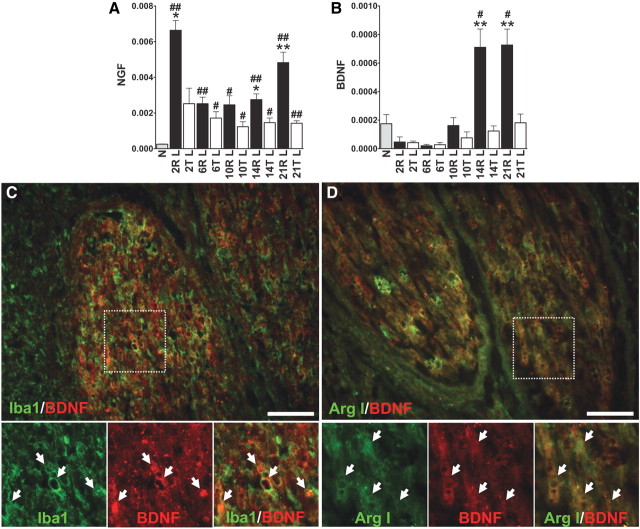
Figure 5.
BDNF and NGF are upregulated in the graft areas of repaired spinal cords. A, NGF expression was upregulated after injury. After 6 d, the expression was maintained at a high level from 2 to 21 d after repair. B, BDNF expression rapidly decreased after injury. After 14 d, expression was significantly induced in the graft areas of repaired spinal cords. #p < 0.05, ##p < 0.01 of group R or T compared with the normal group by Student's t test. *p < 0.05, **p < 0.01 of group R compared with group T by Student's t test. Determinations are means ± SEM from four to five experiments. C, D, Strong immunofluorescence of BDNF was found in the grafted nerves. Many Iba1-positive macrophages (C) and Arg I-positive M2 macrophages (D) in graft areas of 14 d repaired spinal cords colocalized with BDNF immunoreactivity. High-power magnifications of the insets in C and D are shown. Individual double-labeled cells are indicated by arrows. Scale bar, 100 μm.

