Figure 1.
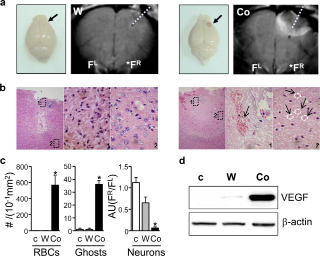
PFC lesions caused by Co-wire lead to increased RBCs, ghost cells, and VEGF expression 5 d after Co-wire implantation. a, The brains of the cobalt-implanted group showed bloody scarring, and MRI displayed that Co-wire implantation led to more profound damage in the PFC than tungsten wire. b, Representative images with hematoxylin/eosin staining of the cobalt- and tungsten-implanted groups. The cobalt group showed increased neuronal death (images 1 and 2 in the Co-wire group) with increased blood cells (an arrow shows RBCs in image 1 in the Co-wire group), and ghost cells (four arrows show the pink ghost cells in image 2 in the Co-wire group), while the tungsten-implanted group showed damaged neuronal cells in limited regions (image 1 in tungsten-implanted group) with morphologically normal neurons in the surrounding area (image 2 in tungsten-implanted group). Left, Ipsilateral PFC (100×); middle, magnified image 1 of ipsilateral PFC (1000×); right, magnified image 2 of ipsilateral PFC (1000×). c, RBCs and ghost cells were significantly increased in cobalt-implanted group. Morphologically normal neurons were largely decreased in Co-wire-implanted group, compared with nonlesion control and tungsten-implanted groups. Arbitrary units (AU) were calculated by dividing the number of neurons in right PFC by the number of neurons in left PFC (FR/FL) d, The level of VEGF (65 kDa) was significantly increased in the cobalt-implanted group. Ghosts, Ghost cells. FR, right prefrontal cortex; FL, left prefrontal cortex. *p < 0.05. Data represent means ± SEM.

