Figure 6.

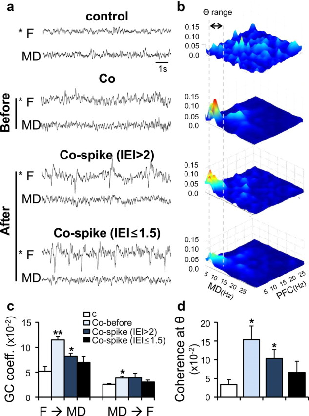
Enhanced thalamocortical resonance was observed before the generation of frontal lobe-specific spikes. a, Representative EEG traces after Co-wire implantation and nonepileptic control. Co-before, 4.2 ± 0.3 d after cobalt implanted; Co-spike (IEI >2), 5.8 ± 0.2 d after cobalt implanted; Co-spike (IEI ≤1.5), 7.6 ± 0.5 d after cobalt implanted. b, Averaged bicoherence analysis between the PFC and MD. Gray dotted lines show the range of theta frequency (4–7 Hz). c, Granger causality analysis between the PFC and MD during development of frontal lobe spikes. d, The comparision of the coherence value at theta range (4–7 Hz) during development of frontal lobe spikes. contr, Contralateral; ipsi, ipsilateral. *p < 0.05, **p < 0.01. Data represent means ± SEM.
