Figure 8.
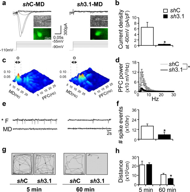
Frontal lobe-specific oscillations and increased thalamocortical interaction at theta range with locomotor hyperactivity were abolished by a region-specific knockdown of the CaV3.1 gene in the MD. a, Lentivirus harboring CaV3.1-specific shRNA led to the reduction of T-type Ca2+ currents in MD neurons. Insets indicate infected neuron visualized with green fluorescence. b, Quantified T-current amplitude in both groups. *p < 0.05. c, Averaged bicoherence analysis between the PFC and MD in shC- and sh3.1-transfected group with Co-wire implantation (4.6 ± 0.2 d after cobalt implanted). Gray dotted lines show the range of theta frequency (4–7 Hz). d, The theta frequency of PFC was also decreased in the sh3.1 group, compared with shC group (4.6 ± 0.2 d after cobalt implanted). e, Representative EEG traces 6 d after Co-wire was implanted. f, The comparision of spike events between the shC/sh3.1 group,6 d after Co-wire implantation. g, The example of locomotor patterns of the sh3.1 group and control group during the first 5 min (5 min) and last 5 min (60 min) of a 60 min recording, 6 d after Co-wire implantation. h, The sh3.1 group showed decreased hyperactivity, compared with shC group (6 d after cobalt wire was implanted). *p < 0.05. Data represent means ± SEM.

