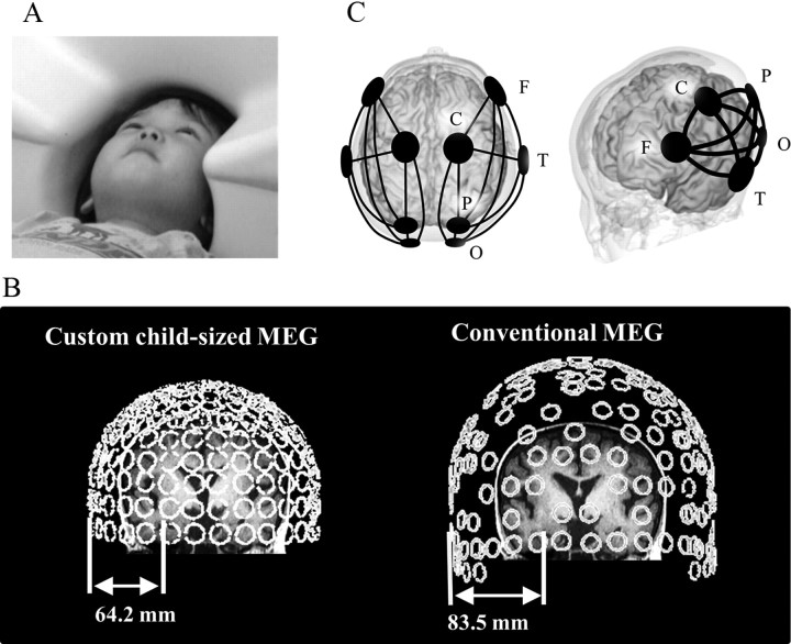
Figure 1.
A, In the custom child-sized MEG system, the MEG sensors are as close to the whole head as possible for optimal recording in young children. During MEG recording, the children lay supine on the bed and viewed video programs projected onto a screen. B, Distances between MEG sensors (temporal area) and hippocampus. In 3- to 4-year-old children (left), sensors were close enough to the parahippocampal gyrus to record its activity, whereas in a conventional MEG system (right), the distance is too great to obtain a good signal from the parahippocampal gyrus. Open circles indicate MEG SQUID sensors (rear view). C, Schema of five selected sensors and 10 connections of interest (solid line) in each hemisphere.

